This one is tricky when isolated, but it is very important not to miss. We treat it just like any other ST segment elevation MI, which is of course time sensitive.
The posterior wall is supplied by the posterior descending artery. The PDA branches from the right coronary artery in 80% of people (those who are right coronary dominant); therefore, occlusion of RCA can result in both an inferior STEMI and a posterior MI as well. Sometimes, it is obvious on the ECG when a posterior MI accompanies an inferior STEMI, but it can also occur all by itself.
The ECG criteria to diagnose a posterior MI — treated like a STEMI, even though no real ST segment elevation is apparent — include:
- ST segment depression (not elevation) in V1 to V4. Think of things backwards. These are the septal and anterior ECG leads. The MI is posterior (opposite to these leads anatomically), so there is ST depression instead of elevation. Turn the ECG upside down, and it would look like a STEMI.
- The ratio of the R wave to the S wave in leads V1 or V2 is greater than 1. This represents an upside-down Q wave (similar in reason to the ST depression instead of elevation).
- ST segment elevation in the posterior leads of a posterior ECG (leads V7-V9). A posterior ECG is done by simply adding three extra precordial leads wrapping around the left chest wall toward the back.
Below are some examples including isolated posterior MIs, inferior STEMIs with posterior involvement and a posterior ECG.
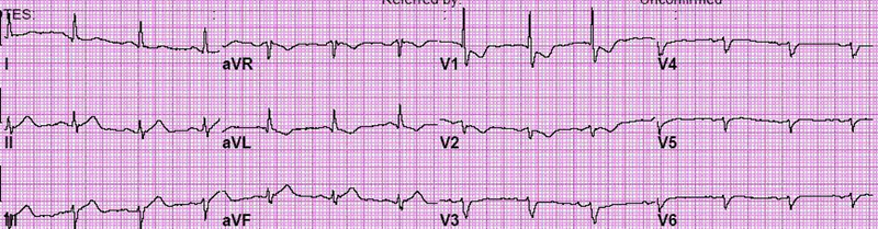
Here is a patient with an isolated posterior MI. There is no inferior involvement here. It would have been nice to see more ST depression in V2, but there is some. Note the R/S ratio in V1 is quite high.
Now, here is the same patient with a posterior ECG tracing. Leads V7 to V9 were added. Leads V1 and V2 were moved a bit just to confuse us. There is not quite 1 mm ST segment elevation in these posterior leads, but you can see at least some slight elevation.

Below are two examples of ECG tracings with both inferior STEMI and posterior involvement. Remember, the more you look at the better!
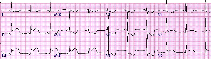
Inferior-Posterior STEMI Example #1
Inferior-Posterior STEMI Example #2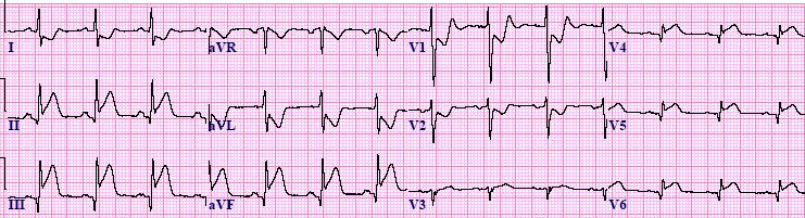
دیدگاه خود را بنویسید